Corneal Conditions
 The cornea is the eye’s outermost layer. It is the clear, domeshaped surface that covers the front of the eye. It plays an important role in focusing your vision.
Cicatricial Pemphigoid


 The ocular cicatricial pemphigoid and Stevens-Johnson Syndrome are conditions associated with severe scarring of the conjunctiva and cornea. These are rare but potentially visually disabling conditions that frequently have multi-system involvement necessitating a multidisciplinary approach for diagnosis and treatment. Oral therapy is frequently required using strong medications that need careful monitoring for side effects. Various surgical techniques are available to try to repair the ocular surface and maintain or improve vision.
Conjunctival Tumor Excision
 Small ocular cancers on the surface of the eye are generally surgically excised. Additional cryotherapy (freezing treatment) may be used.
Conjunctivitis (“Pink Eye”)
 Conjunctivitis, or “pink eye,” is a common condition that often affects school-age children though it also affects adults. It occurs when the conjunctiva (the clear membrane that lines the eyelids and covers the outer edge of the cornea) becomes inflamed from an infection, allergy or irritant. Infectious conjunctivitis is caused by a virus or bacteria from contaminated fingers, towels, washcloths or any other material that touches the eye. Because children interact with each other so much, it is not uncommon for several playmates or schoolmates to come down with infectious conjunctivitis at about the same time. Infectious conjunctivitis causes the white part of the eye (the sclera) to become red and to feel sandy or gritty. If the cause of the infection is bacterial, a yellow pus-like discharge will appear and form a crusty ridge around the eyelid. If it is viral, the discharge is usually be clear and watery. If you suspect your child has infectious conjunctivitis, see your eye care physician immediately. Treatment for bacterial infection involves the washing away of the discharge as well as the use of antibiotics.
  It usually takes one to two weeks for this condition to clear. In addition, you may want to be sure your child does not spread infectious conjunctivitis by insisting that he or she washes his or her hands and does not share washcloths. Viral conjunctivitis frequently resolves on its own. Allergic conjunctivitis affects persons with hayfever or allergies to certain substances or cosmetics. Long-term allergies tend to make the eye red and irritated. With allergic conjunctivitis, there is a watery discharge that accompanies this bothersome condition. During hayfever season, some individuals may experience a short-term white puffiness that affects the conjunctiva. This may last for a few hours, and then clear. The best way to treat allergic conjunctivitis is to locate the cause of the allergy and avoid it. It may also be necessary to take regularly antihistamines to relieve allergy symptoms. Foreign bodies and chemicals may cause another form of conjunctivitis. This condition is nearly always preventable if you remember to wear certified eye protection whenever you are working near hazardous substances or even performing chores like mowing the lawn or sawing word. Click here for tips on how to prevent this and other eye injuries. Most cases of conjunctivitis are not serious. However there is one exception. “Ophthalmia neonatorum” is a type of conjunctivitis that affects newborn babies.
  This sometimes occurs from contact with the lining of the birth canal. This condition is extremely serious and could result in blindness if not treated immediately.
Cornea Edema After Cataract Surgery
 Some patients develop permanent corneal edema (swelling) after cataract surgery. They may have had mild Fuchs’ corneal dystrophy which was aggravated by the cataract surgery. Patients with significant corneal edema may require corneal transplantation to regain good vision.
Corneal Clouding
 Clouding of the cornea, whether from external or internal sources, can mean partial or total loss of sight, depending on the severity of damage. A normal, undamaged cornea is necessary for full vision. When the cornea becomes cloudy, similar to the frosting of glass, there are various methods to correct or restore vision. When the cloudiness is on the surface of the cornea, the top layers can be shaved off with a blade or a laser. When deeper layers of the cornea are involved, it may require replacement. Cornea transplants, using tissue from donated eyes, have been successful at treating this condition. Approximately 40,000 corneal transplants are performed in the United States each year. There are many reasons why the normal, clear cornea may be cloudy, resulting in loss of vision in one of both eyes:
Corneal Topography

 Proper curvature of the cornea is extremely important in bending light to focus on the retina to obtain good vision. Certain conditions cause the corneal curvature to become abnormal, such as keratoconus. A computerized analysis of corneal curvature, called corneal topography, can detect small changes in corneal curvature. This analysis can help determine the cause of decreased vision and often the solution.
Corneal Transplantation
 When the cornea no longer provides good vision because of swelling, scarring, or irregularity, corneal transplantation may be necessary. This procedure involves removing approximately two-thirds of the patient’s cornea and replacing it with a donor cornea. The corneal transplant surgery may be combined with cataract removal or other surgery as needed. The surgery is often successful but it typically takes 12 months or longer to achieve good vision after a corneal transplant.
Descemet’s Stripping Endothelial Keratoplasty
 A new technique being used in some types of cornea transplants means a shorter recovery time for patients and may lead to even better vision after surgery. The advance could make it possible for eye specialists to offer cornea transplants to visually impaired patients in Third World countries and other areas where such procedures have previously been impractical. The technique, known as Descemet’s Stripping Endothelial Keratoplasty (DSEK), allows surgeons to remove a much smaller portion of the patient’s cornea compared with older methods of transplantation. Standard human cornea transplants involve removal of the full thickness of the patient’s cornea and replacement with full thickness donor tissue. In the newer method, surgeons remove only the diseased layer of cells at the back of the cornea and replace it with a similar amount of donor tissue. Doing so helps to retain the structural integrity and mechanical strength of the eye, and typically means a shorter recovery period for the patient. Approximately 35,000 cornea transplants are performed each year in the U.S. Use of the DSEK technique is limited to patients with damage to the endothelial area of the cornea, which accounts for about two-thirds of these cases. The cornea is comprised of several layers, including Descemet’s membrane and the endothelium. Descemet’s membrane is a thin layer that supports endothelial cells on the back of the cornea. The endothelium, comprised of endothelial cells, is the delicate back portion of the cornea that manages fluid content within the corneal structure. In a DSEK procedure, the surgeon removes the inner-most layer of the endothelium along with Descemet’s membrane. That section is then replaced with a delicate healthy layer of donated human cornea tissue. Using specialized tools, the new tissue is positioned into the cornea. The new tissue heals without sutures.
Collagen Crosslinking
 Corneal collagen cross-linking riboflavin therapy is a relatively new experimental treatment for KC. The process works by strengthening the collagan fibers which provide the structural integrity of the cornea. The treatment consists of applying riboflavin (vitamin B2) eyedrops to the corneas and then exposing them to ultraviolet light to activate the riboflavin, allowing it to bind to the collagen in the cornea. Click on the KC Research section of this website to review some of the clinical research on collagen cross-linking therapy. Although it is a relatively new procedure and still being investigated, the hope is that the process will halt the progression of keratoconus.
Corneal Ulcer
 Ulceration of the cornea typically presents with a red, painful eye, with mild to severe discharge and reduced vision. The condition represents a localized infection of the cornea similar to an abscess. Most cases are due to a bacterial infection, commonly associated with antecedent trauma, especially contact lens wear . Other causative agents include fungi, acanthamoeba (a parasite), which typically occurs in contact-lens wearers who swim with the lenses in, or herpes simplex virus infection.
Bacterial Corneal Ulcer
Patients suspected of having a bacterial corneal ulceration are usually treated with frequent application of topical antibiotics , with or without initial cultures. The location and size of the ulceration will guide the ophthalmologist regarding the necessity of cultures. Most patients are followed every one to three days depending on the severity of the condition. If the ulceration is in the central cornea, resolution of the condition is typically slower and vision may be permanently reduced due to scarring, despite early presentation and treatment. 

Fungal Corneal Ulcer
Fungal keratitis (ulceration) may be suspected after trauma with vegetable matter such as a tree branch. In most cases, the eye is already compromised by pre-existing conditions. The diagnosis of fungal keratitis can only be made with microscopic evaluation of specially stained specimens or cultures. Anti-fungal agents are administered, sometimes both topically to the eye and orally, depending on the severity of the ulceration. The prognosis for good vision depends on the extent of infection. Some cases will require corneal transplantation (penetrating keratoplasty) despite early presentation and proper management. 
Dry Eye Syndrome
 Dry eye syndrome is one of the most common problems treated by eye physicians. Over ten million Americans suffer from dry eyes. It is usually caused by a problem with the quality of the tear film that lubricates the eyes. Tears are comprised of three layers. The mucus layer coats the cornea, the eye’s clear outer window, forming a foundation so the tear film can adhere to the eye. The middle aqueous layer provides moisture and supplies oxygen and other important nutrients to the cornea. This layer is made of 98 percent water along with small amounts of salt, proteins and other compounds. The outer lipid layer is an oily film that seals the tear film on the eye and helps to prevent evaporation.
  Tears are formed in several glands around the eye. The water layer is produced in the lacrimal gland, located under the upper eyelid. Several smaller glands in the lids make the oil and mucus layers. With each blink, the eyelids spread the tears over the eye. Excess tears flow into two tiny drainage ducts in the corner of the eye by the nose. These ducts lead to tiny canals that connect to the nasal passage. The connection between the tear ducts and the nasal passage is the reason that crying causes a runny nose.
  In addition to lubricating the eye, tears are also produced as a reflex response to outside stimulus such as an injury or emotion. However, reflex tears do little to soothe a dry eye, which is why someone with watery eyes may still complain of irritation. Dry eye syndrome has many causes. One of the most common reasons for dryness is simply the normal aging process. As we grow older, our bodies produce less oil – 60% less at age 65 then at age 18. This is more pronounced in women, who tend to have drier skin then men. The oil deficiency also affects the tear film. Without as much oil to seal the watery layer, the tear film evaporates much faster, leaving dry areas on the cornea. Many other factors, such as hot, dry or windy climates, high altitudes, air-conditioning and cigarette smoke also cause dry eyes. Many people also find their eyes become irritated when reading or working on a computer. Stopping periodically to rest and blink keeps the eyes more comfortable. Contact lens wearers may also suffer from dryness because the contacts absorb the tear film, causing proteins to form on the surface of the lens. Certain medications, thyroid conditions, vitamin A deficiency, and diseases such as Parkinson’s and Sjogren’s can also cause dryness. Women frequently experience problems with dry eyes as they enter menopause because of hormonal changes. Symptoms -Itching -Burning -Irritation -Redness -Blurred vision that improves with blinking -Excessive tearing -Increased discomfort after periods of reading, watching TV, or working on a computer
Detection and Diagnosis
There are several methods to test for dry eyes. The doctor will first determine the underlying cause by measuring the production, evaporation rate and quality of the tear film. Special drops that highlight problems that would be otherwise invisible are particularly helpful to diagnose the presence and extent of the dryness. Your ophthalmologist is often able to diagnose dry eye by simply examining the eyes. Sometimes tests that measure tear production may be necessary. Such tests may involve placing filter-paper strips under the lower eyelids to measure the rate of tear production under various conditions. 
Treatment
Your ophthalmologist is often able to diagnose dry eye by simply examining the eyes. Sometimes tests that measure tear production may be necessary. Such tests may involve placing filter-paper strips under the lower eyelids to measure the rate of tear production under various conditions. Dry eye is sometimes treated using eye drops called “artificial tears.” Artificial tears are available without a prescription. You may want to try several brands to find the one that you like best. Preservative-free eye drops are available if you are sensitive to the preservatives in artificial tears. If you need to use artificial tears more than every two hours, preservative-free brands may be better for you. Solid lid inserts that gradually release lubricants during the day may also be beneficial for some people. Some patients with mild dryness may benefit from blinking more frequently, especially when reading, driving, or watching television. Conserving the tears in your own eyes is another approach to keeping the eyes moist. Tears drain out of the eye through a small canal into the nose (that is why your nose runs when you cry). Your ophthalmologist may close these canals either temporarily or permanently. This closure conserves your own tears and makes artificial tears last longer. Other methods may include steps to prevent the evaporation of tears. In winter, when indoor heat is on, a humidifier or a pan of water on the radiator will add moisture to dry air. Wrap-around glasses (illegal to wear while driving in some states) may reduce the drying effect of the wind. Anything that adds to dryness such as an overly warm room, hair dryers, or wind should be avoided by a person with dry eye. Smoking is especially bothersome. Some people with dry eye complain of “scratchy eyes” upon awakening. This symptom can be treated by using an ointment at bedtime. Use the smallest amount of ointment necessary for comfort, since the ointment can cause your vision to blur temporarily.  Dry eye caused by a lack of vitamin A in the diet is rare in the United States, but is more common in poorer countries. Ointments containing vitamin A can help dry eye caused by this unusual condition. Vitamin A does not seem to help people with ordinary dry eye.
Dystrophies
 Acorneal dystrophy is an abnormality of a specific part of the cornea that leads to corneal clouding. It may occur early in life, but is usually seen between the ages of 20 and 50. The dystrophy most commonly requiring a corneal transplant is Fuchs’ dystrophy, a condition often found in older adults.
Excimer Laser Phototherapeutic Keratectomy

 The excimer laser (the same laser used for refractive surgery) can also be used to remove opacities from the surface of the cornea. Patients with corneal dystrophies affecting the front of the cornea, such as granular and lattice dystrophies, or with superficial
Herpes Simplex Keratitis

 The herpes simplex virus, the same virus that causes cold sores around the mouth (not the one that causes venereal disease), can infect the cornea and cause pain and decreased vision. Treatment is with antiviral and often anti-inflammatory medications. Occasionally, severe corneal scarring can be treated with surgery.
Keratoconus


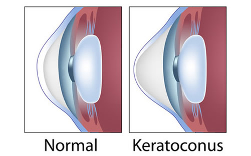
 Keratoconus is a disease which causes the cornea to protrude and become progressively thinner and cone-shaped. Eyeglasses usually cannot fully correct this condition, and in advanced cases, patients can only obtain useful vision with specially designed hard lenses. Very severe cases that cannot be fit with lenses or cannot get useful vision with contact lenses may require a corneal transplant.
Pterygium


 Apterygium is an excessive growth of scar-like tissue onto the surface of the cornea. It can cause redness and irritation. An advanced case of pterygia can affect vision by causing corneal irregularity or by growing over the center of the cornea, but it can be treated with surgery.
Recurrent Erosion Syndrome

 Recurrent painful episodes typically result from previous corneal abrasions but may occur spontaneously in patients with predisposed abnormal corneas. The episodes generally occur at night or upon awakening in the morning. This condition results from a breakdown of the surface layer of the cornea causing a painful “scratched” cornea. Treatment options include medications and minor surgical procedures.
Specular Microscopy

 The endothelial cells on the back surface of the cornea pump fluid out of the cornea to keep it clear. When these cells do not function properly, the cornea swells, the vision decreases, and pain can develop. Specular microscopy can visualize the endothelial cells to help determine their health.